Quick answer: A urinalysis is a three-part urine test that checks the physical appearance of your urine, runs a chemical dipstick across 10 to 12 parameters (protein, glucose, blood, leukocytes, nitrites, pH, specific gravity, ketones, bilirubin, urobilinogen), and, when ordered, adds a microscopic exam of spun sediment to count cells and crystals. It takes about 30 minutes in most labs and catches early-stage kidney disease, urinary tract infections, diabetes, and several metabolic problems before symptoms appear. Normal results show no protein, no glucose, no blood, no nitrites, and a pH between 4.5 and 8.0, though reference ranges vary slightly by lab.
What does a urinalysis actually measure?
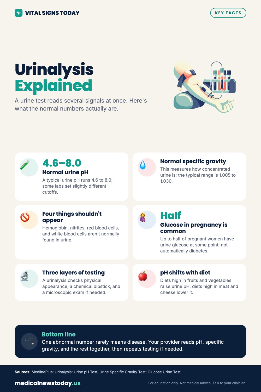
A standard urinalysis measures three separate layers of information from a single urine sample. Most people see only the final report, which makes the results feel like a random assortment of words and plusses, but the logic is straightforward once you understand the three components.
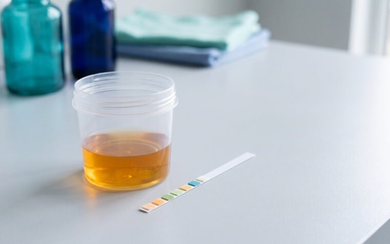
The physical exam is what a technician observes before any chemistry: color (pale yellow to deep amber), clarity (clear versus cloudy or turbid), and sometimes odor if it is unusual. Cloudy urine usually means white cells, bacteria, or crystals. Orange urine can mean dehydration, bilirubin, or certain antibiotics. This step takes about 30 seconds and can flag problems before a single reagent touches the sample.
The chemical dipstick exam is the workhorse. A plastic strip with 10 or more small reagent pads is dipped into the urine for 60 seconds. Each pad changes color in proportion to the concentration of its target analyte. Lab readers scan the strip optically to reduce technician subjectivity. This is where protein, glucose, ketones, blood, leukocyte esterase, nitrites, pH, specific gravity, bilirubin, and urobilinogen are detected.
The microscopic exam is performed when the dipstick flags something abnormal, or when the ordering clinician requests it. The urine is centrifuged, the sediment is placed on a slide, and a technician counts red blood cells (RBCs), white blood cells (WBCs), casts (cylindrical molds formed in the kidney tubules), epithelial cells, crystals, and bacteria per high-power field. This is the most informative and most expensive part of a complete urinalysis, and it is the step that separates a real kidney problem from a dipstick artifact. For a broader look at what labs routinely screen, see the best biomarkers to test.
What are normal urinalysis results?
Normal urinalysis results have fairly consistent ranges across Quest Diagnostics and Labcorp, though exact cutoffs differ slightly between labs and instruments. The table below reflects the most widely accepted reference intervals for adults.
| Parameter | Normal range | What a positive or abnormal result suggests |
|---|---|---|
| Color | Pale to dark yellow | Orange (bilirubin, rifampin), red/pink (blood, beets, certain meds), green (Pseudomonas, propofol) |
| Clarity | Clear to slightly hazy | Cloudy: pyuria, bacteriuria, phosphate crystals |
| pH | 4.5 to 8.0 | Very alkaline (>8): UTI with urease-producing bacteria, renal tubular acidosis; very acidic (<4.5): high-protein diet, metabolic acidosis |
| Specific gravity | 1.003 to 1.035 | <1.003: dilute (excessive hydration, diabetes insipidus); fixed at 1.010: severe kidney disease |
| Protein | Negative (<8 mg/dL) | Trace to 3+: kidney disease, pre-eclampsia, multiple myeloma, intense exercise |
| Glucose | Negative | Positive: blood glucose >180 mg/dL (diabetes), SGLT2 inhibitor use, renal glycosuria |
| Ketones | Negative | Positive: diabetic ketoacidosis, prolonged fasting, very low-carb diet |
| Blood | Negative | Trace to 3+: UTI, kidney stones, trauma, glomerulonephritis, bladder cancer risk factor if persistent |
| Leukocyte esterase | Negative | Positive: WBCs present, suggests infection or inflammation |
| Nitrites | Negative | Positive: gram-negative bacteria converting dietary nitrates (strong UTI indicator) |
| Bilirubin | Negative | Positive: liver disease, bile duct obstruction |
| Urobilinogen | 0.2 to 1.0 EU/dL | Elevated: hemolytic anemia, liver disease; absent: complete bile duct blockage |
| RBCs (microscopic) | 0 to 2 per high-power field | >3: hematuria, requires further workup |
| WBCs (microscopic) | 0 to 5 per high-power field | >5: pyuria, UTI, or sterile inflammation |
| Casts | Hyaline casts only (0 to 5) | RBC casts: glomerulonephritis; WBC casts: pyelonephritis; granular casts: chronic kidney disease |
One caveat worth knowing: a single abnormal dipstick finding rarely closes the case. Leukocyte esterase without nitrites is common after exercise or in women due to vaginal contamination. Protein that disappears after rest is called orthostatic proteinuria and is benign in young adults. Repeating a suspicious result on a first-morning, midstream, clean-catch sample eliminates most artifacts before anyone orders a biopsy.
What does protein in urine mean?
Protein in urine, called proteinuria, is one of the most clinically important urinalysis findings because healthy kidneys filter almost all protein back into the bloodstream. If protein leaks through the glomerular filtration barrier, something is damaging it.
A dipstick reads protein on a scale of negative, trace, 1+, 2+, and 3+. Trace to 1+ is worth repeating but not immediately alarming. Persistent 1+ or higher deserves a spot urine protein-to-creatinine ratio and, if that is elevated, a referral to nephrology. The albumin test is a more sensitive companion: a urine microalbumin-to-creatinine ratio catches early diabetic kidney disease years before a dipstick becomes positive.
Common causes ranked by clinical frequency:
- Diabetic nephropathy (most common cause of chronic proteinuria in the US)
- Hypertensive nephropathy
- Glomerulonephritis (IgA nephropathy, lupus nephritis)
- Orthostatic proteinuria (benign, only in upright position, common in teens)
- Pre-eclampsia (in pregnancy, a medical emergency)
- Multiple myeloma (produces Bence Jones proteins, often missed by dipstick)
- Intense exercise (resolves within 24 to 48 hours)
- Fever
The insider fact most patients do not hear: dipstick proteinuria primarily detects albumin, so patients with myeloma producing immunoglobulin light chains can have heavy protein excretion and a falsely negative dipstick. If myeloma is suspected, a urine protein electrophoresis is required, not just a dipstick.
What causes blood in urine?
Blood in urine, or hematuria, is detected on dipstick by a pad sensitive to hemoglobin and myoglobin, not intact red blood cells. That distinction matters because myoglobinuria (from rhabdomyolysis or extreme exercise) will read positive on dipstick but show zero RBCs on microscopy. True hematuria requires both a positive dipstick and confirmed RBCs on microscopic exam.
Gross hematuria (visible pink or red urine) is always an indication to call a clinician the same day. Microscopic hematuria (more than 3 RBCs per high-power field on two of three properly collected specimens) is present in about 2 to 3 percent of the general adult population at any given time. The cause is benign in the majority of cases, but persistent unexplained microscopic hematuria in adults over 35 requires cystoscopy to rule out bladder cancer, because bladder cancer is notoriously asymptomatic until advanced.
Common causes:
- Urinary tract infections (most common cause overall)
- Kidney stones (often flank pain, but not always)
- Benign prostatic hyperplasia (BPH) in men over 50
- Vigorous exercise, especially long-distance running (clears within 72 hours)
- Anticoagulant therapy (warfarin, rivaroxaban) lowering the threshold for micro-bleeding
- Glomerulonephritis (dysmorphic RBCs or RBC casts on microscopy point here)
- Bladder or kidney cancer (age and smoking are the key risk factors)
- Trauma, recent Foley catheter placement
Menstrual contamination is the most common benign cause of a positive dipstick blood in women. A clean-catch midstream collection taken two days after the last day of menstruation eliminates most of this artifact.
What does glucose in urine mean?
Glucose normally appears in urine only when blood glucose exceeds roughly 180 mg/dL, the threshold at which the kidneys can no longer reabsorb it all, called the renal threshold for glucose. Finding glucose on a urinalysis therefore points strongly toward uncontrolled diabetes, or previously undiagnosed diabetes, and warrants a fasting glucose and HbA1c the same week.
Two situations break this rule. First, SGLT2 inhibitors (empagliflozin, dapagliflozin, canagliflozin) are prescribed to deliberately dump glucose into the urine as a mechanism of action. Any patient on one of these drugs will have persistent glycosuria on dipstick; it is expected and not a diagnostic finding. Second, a rare inherited condition called renal glycosuria lowers the renal threshold, so glucose spills at normal blood sugar. These patients have a normal HbA1c and a positive urine glucose for life.
Important for anyone using a urinalysis to monitor diabetes: urine glucose is a delayed, insensitive marker compared to a blood glucose meter or a continuous glucose monitor. It tells you something happened, not what is happening now. A complete blood panel that includes fasting glucose is far more actionable for diabetes management than following urine glucose.
What do leukocytes in urine mean?
Leukocytes, specifically white blood cells, in urine signal inflammation or infection somewhere in the urinary tract. The dipstick detects leukocyte esterase, an enzyme released by WBCs, which is a useful proxy but not a direct cell count. The microscopic exam provides the actual count.
More than 5 WBCs per high-power field is called pyuria. The combination of positive leukocyte esterase and positive nitrites on dipstick has a sensitivity of about 75 percent and specificity of about 82 percent for a UTI, which is good enough to start empirical treatment in a symptomatic woman without waiting for culture results. In a man, a child, or anyone with recurrent UTIs, a urine culture is mandatory to identify the organism and its antibiotic sensitivities before treatment.
Sterile pyuria, meaning WBCs present but culture negative, is a clinically important finding. Causes include:
- Partially treated bacterial UTI
- Urethritis from Chlamydia trachomatis or Neisseria gonorrhoeae (neither grows on standard urine culture)
- Renal tuberculosis (a classic cause, now rare in the US but still seen in immigrant populations)
- Interstitial nephritis (often drug-induced, especially NSAIDs and proton pump inhibitors)
- Bladder cancer
WBC casts on microscopy are the critical distinguishing finding: if WBC casts are present, the inflammation is originating inside the kidney tubules, not just in the bladder. That finding points toward pyelonephritis or interstitial nephritis and changes management significantly.
How urinalysis fits into a complete health workup
A urinalysis alone is an incomplete picture. It tells you about kidney filtration function and lower urinary tract health, but it says nothing about serum creatinine, eGFR, electrolytes, glucose in the blood, liver enzymes, or lipids. A urinalysis showing early proteinuria should be paired with an albumin test (urine microalbumin-to-creatinine ratio) and a serum creatinine for a meaningful kidney health assessment.
Annual physicals at most US primary care offices include a urinalysis, a basic metabolic panel, and a complete blood count as baseline screening. If you want a truly comprehensive metabolic snapshot, reviewing the best biomarkers to test gives context for where urinalysis fits in relation to lipids, inflammation markers, thyroid, and hormones.
Urinalysis results are also affected by how the sample is collected. A midstream clean-catch into a sterile cup is standard. First morning urine is more concentrated and more likely to show protein, cells, and casts if they are present, making it the preferred specimen for kidney disease screening. Random daytime urine is fine for most clinical purposes but less sensitive for low-level proteinuria.
The simplest way to actually get this done
Superpower is a full-body lab membership that runs 100+ biomarkers, has each result reviewed by a doctor, and tracks your numbers year over year (about $199/year). It is what we point readers to when they would rather get one clean, complete draw than chase single tests one at a time. Here is superpower blood test reviewed in full.
How much does a urinalysis cost?
A standalone urinalysis is one of the cheapest diagnostic tests in outpatient medicine. Prices vary significantly by setting.
| Setting | Estimated cash price (2026) | Notes |
|---|---|---|
| Quest Diagnostics (self-pay portal) | $11 to $29 | Dipstick only; microscopic adds $10 to $25 extra |
| Labcorp (self-pay) | $12 to $35 | Complete UA with microscopy closer to $30 to $55 |
| CVS MinuteClinic / urgent care | $40 to $90 | Includes provider evaluation; insurance often covers UTI visits |
| Primary care office with insurance | $0 to $30 copay | Preventive visit urinalysis usually covered; standalone visit varies |
| Emergency department | $150 to $400+ | Bundled with facility fees; avoid for routine screening |
| HSA/FSA eligible | Yes, all settings | Use pre-tax dollars to bring effective cost down further |
Medicare Part B covers urinalysis when medically necessary, which includes monitoring known kidney disease or diabetes. It is not covered as an isolated screening test under Medicare without a clinical indication. Medicaid coverage rules vary by state but generally mirror Medicare for medically necessary urinalysis.
At-home dipstick strips (brands like Rapid Response and AZO Test Strips) sell at CVS and Amazon for $12 to $20 for 50 strips and check 10 parameters reasonably well for home UTI monitoring. They do not perform microscopy, so they miss casts and cellular detail. They are useful for tracking a known condition between clinic visits, not for replacing a laboratory specimen.
What clinicians look for on a urinalysis that patients miss
The findings that generate the most clinical meaning are not always the ones patients notice on the report. Three examples:
Casts. Most patients skip the microscopy section entirely. Cast type tells a nephrologist more about kidney pathology than almost any other single urinalysis finding. Red blood cell casts are pathognomonic (specific by definition) for glomerulonephritis and require urgent nephrology referral. Granular casts, sometimes called muddy brown casts, are the hallmark of acute tubular necrosis, which is the most common cause of hospital-acquired acute kidney injury. A normal urinalysis in someone whose creatinine is rising sharply is itself useful information because it makes prerenal (dehydration or reduced blood flow) more likely than intrinsic renal damage.
Specific gravity tells you about hydration AND tubular function. A specific gravity above 1.025 in someone with rising creatinine says the kidneys are trying to conserve water, which points toward dehydration or heart failure reducing renal perfusion. A specific gravity fixed at 1.010 regardless of fluid intake means the tubules have lost their concentrating ability, a marker of chronic kidney disease or acute tubular necrosis. Clinicians read this number before they read creatinine.
Urobilinogen elevation without bilirubin. This pattern, elevated urobilinogen but negative bilirubin, suggests hemolytic anemia or early hepatocellular liver disease rather than obstructive jaundice. Obstructive jaundice (a blocked bile duct) produces bilirubin in the urine without elevated urobilinogen because bilirubin cannot reach the gut to be converted. The pattern of the two together is more diagnostic than either alone. For liver-related workup, an alkaline phosphatase test is a natural companion.
FAQ
What is a urinalysis used to diagnose?
A urinalysis is used to diagnose or screen for urinary tract infections, kidney disease (glomerulonephritis, diabetic nephropathy, chronic kidney disease), diabetes and prediabetes (through glucose and ketones), liver disease (bilirubin, urobilinogen), and dehydration. It is also part of pre-operative screening and routine prenatal care. A single abnormal result rarely confirms a diagnosis; it guides the next test.
Do I need to fast before a urinalysis?
No fasting is required for a standard urinalysis. You can eat and drink normally before the test. However, first-morning urine, collected before eating or drinking anything, is the most concentrated specimen and is preferred for kidney disease screening and for detecting protein or casts at the lowest possible threshold. If your clinician asks for a first-morning sample, collect it before your first cup of coffee or water.
What does a trace of protein in urine mean?
Trace protein on a dipstick (less than 30 mg/dL) is often insignificant and can result from dehydration, exercise in the preceding 24 to 48 hours, fever, or the simple fact that the urine is concentrated that morning. A single trace result in an otherwise healthy person without diabetes, hypertension, or kidney disease is usually repeated in four to six weeks. If it persists, a urine microalbumin-to-creatinine ratio and serum creatinine clarify whether the kidney filtration barrier is leaking. Trace protein that vanishes on repeat testing does not require treatment.
Can a urinalysis detect sexually transmitted infections?
A standard urinalysis cannot diagnose chlamydia, gonorrhea, or other STIs. It can detect the downstream effects of untreated STIs, specifically sterile pyuria (white cells without bacteria on culture) in someone with urethritis from chlamydia or gonorrhea. Definitive STI diagnosis requires nucleic acid amplification tests (NAAT) run on a urine sample specifically labeled for STI testing, which is an entirely different assay from a urinalysis. Asking your provider for both is common at sexual health visits.
What does it mean if my urinalysis shows leukocytes but no nitrites?
Leukocytes present without nitrites means white blood cells are in the urine but there is no evidence of gram-negative bacteria. This pattern fits several scenarios: a gram-positive UTI like Enterococcus (which does not reduce nitrates to nitrites), an STI-related urethritis, a specimen that sat too long before processing (WBCs lyse), interstitial nephritis, or vaginal contamination in women. A urine culture with the same sample usually resolves the question. About 25 percent of true bacterial UTIs are caused by organisms that do not produce nitrites, so a negative nitrite does not rule out infection.
How is a urinalysis different from a urine culture?
A urinalysis identifies what is in the urine using dipstick chemistry and microscopy, and returns results in under an hour. A urine culture takes 24 to 72 hours and identifies the specific bacteria causing an infection, along with which antibiotics will kill it. They serve different roles: urinalysis flags that an infection is likely, and culture confirms the organism and directs antibiotic selection. Most guidelines recommend a culture before treating complicated UTIs (men, pregnant women, recurrent infections, immunocompromised patients), while uncomplicated UTIs in otherwise healthy young women can often be treated empirically after a positive dipstick alone.
Is a urinalysis the same as a drug test?
No. A clinical urinalysis and a urine drug screen are completely different tests that happen to use the same specimen type. A clinical urinalysis looks at chemical and cellular markers of health. A urine drug screen uses immunoassay to detect metabolites of specific controlled substances. The two tests are ordered separately, processed differently, and reported through different channels. A clinical urinalysis will not reveal recreational drug use, and a drug screen will not diagnose a UTI.
What does a urinalysis show that blood tests miss?
A urinalysis uniquely reveals the composition of the kidney filtrate and the cellular contents of the urinary tract, information blood tests cannot provide. Casts, which form only inside kidney tubules, are invisible on any blood panel. Bacteria and WBCs in the bladder are not reflected in serum white blood cell counts in most uncomplicated UTIs. Bilirubin reaching the urine appears there before it is detectable by symptoms. The superpower blood test review covers how urinalysis and serum biomarkers complement each other in a complete metabolic snapshot. For a broader overview of what to include in routine labs, the adiponectin test is an example of a metabolic marker that pairs well with urine findings in insulin-resistant patients.