Disclosure: Vital Signs Today may earn a commission if you buy through links on this page, at no extra cost to you. This does not influence our evidence-based assessments. We are not a medical provider; talk to a clinician before acting on test results.
Quick answer: RBC indices are four numbers on a standard CBC that describe the size, hemoglobin content, hemoglobin concentration, and size variability of your red blood cells: MCV, MCH, MCHC, and RDW. Clinicians use them to classify anemia into microcytic (small cells, usually iron deficiency or thalassemia), normocytic (normal-sized cells, often chronic disease or blood loss), or macrocytic (large cells, typically B12/folate deficiency or liver disease). An abnormal RBC index does not diagnose a condition on its own, but it tells you exactly which direction to investigate next.
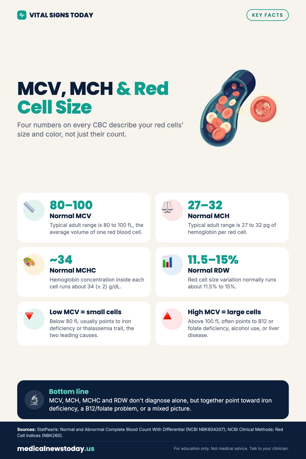
What Are RBC Indices and Why Do They Appear on Every CBC?
RBC indices are a group of calculated and directly measured values that characterize individual red blood cells rather than just counting them. Every complete blood panel ordered at Quest, Labcorp, or any hospital lab automatically generates these numbers from the same blood draw. Modern hematology analyzers measure each red cell electronically and compute the indices in seconds, which is why they add zero cost or extra tubes to a routine CBC.
The four standard indices are:
- MCV (Mean Corpuscular Volume): average volume of a single red blood cell, expressed in femtoliters (fL).
- MCH (Mean Corpuscular Hemoglobin): average mass of hemoglobin per red cell, in picograms (pg).
- MCHC (Mean Corpuscular Hemoglobin Concentration): average concentration of hemoglobin inside each cell, in grams per deciliter (g/dL).
- RDW (Red Cell Distribution Width): a statistical measure of how much variation exists in red cell size, expressed as a coefficient of variation (CV) percentage.
MCH and MCHC are closely tied to MCV because a bigger cell generally holds more hemoglobin. When MCV is low, MCH usually drops with it. MCHC is the most independent of the four; it specifically tells you whether cells are pale (hypochromic) or normally colored (normochromic). RDW is increasingly recognized as a marker of systemic inflammation and nutritional deficiency even before the other indices shift.
MCV Normal Range: What the Numbers Actually Mean
The MCV normal range in most US labs is 80 to 100 fL, though some labs use 80 to 96 fL. A value below 80 fL is microcytic; above 100 fL is macrocytic. A single fL deviation rarely matters clinically, but a sustained MCV of 72 fL or 112 fL points to a real pathology that needs follow-up.
| Index | Normal Range (US labs) | Unit |
|---|---|---|
| MCV | 80 to 100 | fL |
| MCH | 27 to 33 | pg |
| MCHC | 32 to 36 | g/dL |
| RDW-CV | 11.5 to 14.5 | % |
Want to check your red blood cells?
Three ways to get it measured. Pick whichever fits you.
Affiliate links: we may earn a commission at no extra cost to you. A lab test does not replace a visit with your clinician.
Reference ranges differ slightly by laboratory and by the analyzer used. Your report always lists the lab-specific reference interval next to your value, so use that column rather than memorizing generic cutoffs.
One thing most patients do not know: MCV is a mean, so it averages all your red cells together. If you have two simultaneous deficiencies (iron plus B12, for example), small cells and large cells can coexist and produce a falsely normal MCV while your RDW is extremely high. A clinician who looks only at MCV in that scenario will miss a mixed deficiency entirely. That is exactly why RDW exists.
High MCV Causes: When Your Red Cells Are Too Big
A high MCV, called macrocytosis, means red blood cells are larger than normal. The most common causes in the US are B12 deficiency, folate deficiency, alcohol use disorder, liver disease, and certain medications, especially hydroxyurea, methotrexate, and antiretrovirals for HIV.
Breaking it down by mechanism:
- Megaloblastic anemia (B12 or folate deficiency): impaired DNA synthesis forces cells to grow larger before they can divide. The cells come out oversized and often misshapen. A peripheral blood smear will show hypersegmented neutrophils, a near-pathognomonic finding.
- Alcohol: ethanol is directly toxic to red cell precursors in the bone marrow, independent of folate status. Many people with alcohol-related macrocytosis have a normal folate level. MCV can take months to normalize after stopping alcohol because red cells live roughly 120 days.
- Hypothyroidism: slowed metabolism reduces erythropoiesis efficiency, producing larger, slower-maturing cells. This is a frequently missed cause; always check TSH when MCV is elevated without an obvious explanation.
- Liver disease: abnormal lipid deposition alters the red cell membrane and increases surface area, causing a non-megaloblastic macrocytosis. The cells look round and enlarged on smear rather than oval.
- Medications: folate antagonists (methotrexate, trimethoprim) block the same DNA synthesis pathway as dietary deficiency. Hydroxyurea used for sickle cell disease or polycythemia vera routinely raises MCV to 110 to 120 fL as an expected effect.
An MCV above 115 fL almost always indicates megaloblastic change (B12/folate) or a drug effect. Values between 100 and 110 fL are more likely to reflect alcohol, hypothyroidism, or liver disease.
Low MCV Meaning: The Iron and Thalassemia Split
A low MCV, called microcytosis, means red cells are smaller than normal. In US primary care, iron deficiency anemia is responsible for roughly 70 to 80 percent of microcytic cases. Thalassemia trait accounts for most of the rest, followed by anemia of chronic disease (which is usually normocytic but can be mildly microcytic) and sideroblastic anemia.
The clinical trick to separate iron deficiency from thalassemia trait is the Mentzer index: divide MCV by the RBC count. A Mentzer index below 13 suggests thalassemia; above 13 suggests iron deficiency. It is not definitive, but it costs nothing and guides whether to order hemoglobin electrophoresis before loading someone with iron supplements.
Distinguishing features at a glance:
- Iron deficiency: low MCV, low MCH, low MCHC (pale cells), high RDW (because new cells form as iron is depleted, creating a population of tiny pale cells alongside older normal cells), low ferritin, low serum iron, high TIBC.
- Beta-thalassemia trait: low MCV (often dramatically low, 65 to 72 fL), normal or slightly low MCH, normal MCHC, normal or slightly elevated RDW, normal ferritin. The RBC count is typically elevated or normal despite low MCV, because the marrow compensates by making more cells.
- Anemia of chronic disease: MCV low-normal to mildly low, normal or low MCHC, normal RDW, elevated ferritin, low serum iron, low TIBC.
Iron supplementing someone with thalassemia trait causes iron overload without any benefit. Getting the correct diagnosis first is not optional.
MCH vs MCV: They Are Related but Not Interchangeable
MCH (hemoglobin mass per cell) and MCV (volume per cell) move together in most anemias, but they carry different clinical weight. MCV reflects cell size driven by the production process. MCH reflects how much hemoglobin actually ended up packed inside that cell once it matured.
In practice, MCH is often more stable and slightly more sensitive as an early marker of iron deficiency than MCV. Iron deficiency reduces hemoglobin synthesis before it visibly shrinks cell volume, so MCH may fall first. An MCH below 27 pg with a still-normal MCV is a soft early signal worth noting, especially in menstruating women or frequent blood donors.
MCHC is the most specific of the three for hypochromia. A persistently low MCHC (below 31 g/dL) with low MCV and MCH is classic iron deficiency or thalassemia. A high MCHC (above 36 g/dL) is unusual and suggests hereditary spherocytosis, where cells are small and dense. Automated analyzers generate a MCHC above 36.5 g/dL as a quality flag because hemolysis in the tube can falsely elevate it; a lab may reflex to a manual smear review.
If you are trying to decide whether your anemia is from iron deficiency, the MCH and MCHC together are more discriminating than MCV alone. Tracking all three over time after starting iron therapy shows whether the marrow is responding correctly: MCV and MCH should rise toward normal over six to eight weeks as new, properly hemoglobinized cells replace the deficient ones.
RDW Blood Test: The Overlooked Index That Reveals Mixed Deficiencies
RDW measures anisocytosis, the technical term for variability in red cell size. A high RDW means your red cells come in wildly different sizes rather than a uniform population. A low RDW is almost never clinically significant on its own.
RDW is most useful in two situations:
- Unmasking a mixed deficiency: as described above, simultaneous iron deficiency (pushing MCV down) and B12 or folate deficiency (pushing MCV up) can produce a normal or near-normal MCV with a high RDW. The RDW spikes because the population is bimodal, half tiny and half huge. Missing this means treating only one deficiency while the other continues destroying red cells.
- Early iron deficiency before frank anemia develops: RDW rises before MCV falls and before hemoglobin drops below the anemia threshold. A person with ferritin of 8 ng/mL and a hemoglobin of 12.5 g/dL (technically above the anemia cutoff) may already have an RDW of 15.5 percent, flagging the problem early.
Beyond hematology, a growing body of evidence links elevated RDW to cardiovascular disease risk, inflammation, and all-cause mortality, even in people without anemia. Whether RDW causes worse outcomes or simply reflects underlying disease burden is still debated, but seeing an RDW of 17 percent in a 45-year-old who looks healthy on paper should at least prompt a closer look at the full picture. Talk to a clinician about your results if your RDW is persistently above 15 percent.
When you are deciding which biomarkers to track for a comprehensive baseline, RDW belongs on that list as firmly as hemoglobin. See our guide to the best biomarkers to test for a fuller ranking.
How RBC Indices Classify Anemia: The Three-Box System
The most practical use of RBC indices is categorizing any anemia into one of three morphological boxes, each pointing to a different diagnostic workup.
| Category | MCV | Key Causes | Next Tests |
|---|---|---|---|
| Microcytic anemia | <80 fL | Iron deficiency, thalassemia, anemia of chronic disease | Ferritin, serum iron, TIBC, hemoglobin electrophoresis |
| Normocytic anemia | 80 to 100 fL | Acute blood loss, chronic kidney disease, hypothyroidism, hemolysis, aplastic anemia | Reticulocyte count, creatinine, TSH, LDH, haptoglobin |
| Macrocytic anemia | >100 fL | B12 deficiency, folate deficiency, alcohol, liver disease, medications | B12, folate, LFTs, TSH, peripheral smear, medication review |
This three-box approach is taught in every US medical school and used daily in emergency departments, primary care offices, and hematology clinics. What most patients do not realize is that getting to the right box immediately narrows the differential diagnosis from dozens of possible causes to three to five. That is the power of RBC indices: they do not diagnose, but they eliminate most possibilities in seconds.
Normocytic anemia is the trickiest box because it contains the most heterogeneous diseases. The reticulocyte count becomes essential here. A high reticulocyte count in a normocytic anemia points to hemolysis or blood loss (the marrow is producing rapidly). A low reticulocyte count points to underproduction, as in chronic kidney disease or aplastic anemia.
If you are getting blood drawn anyway, it is often smarter to capture a full baseline at once. Here is how a full-body panel compares to piecemeal single-marker testing.
What Causes Abnormal RBC Indices Without Anemia?
You can have abnormal MCV or RDW with a hemoglobin that is technically in the normal range. This is more common than most patients expect and clinically significant in several scenarios.
Macrocytosis without anemia is the most common scenario. An MCV of 104 fL in someone with a normal hemoglobin of 14 g/dL almost always means alcohol use or early B12 deficiency. The marrow is compensating well enough to maintain red cell counts, but the underlying nutritional or toxic problem is present. Left untreated, B12 deficiency will progress to neurological damage (subacute combined degeneration of the cord) even without ever developing overt anemia.
Microcytosis without anemia is the hallmark of thalassemia trait. Someone with alpha or beta thalassemia trait typically has an MCV of 65 to 75 fL and a normal hemoglobin because the marrow compensates by producing extra cells. Many people carry thalassemia trait their entire lives without knowing, and it matters mainly for genetic counseling (two carriers have a 25 percent chance of a child with thalassemia major).
Elevated RDW without anemia can reflect early nutritional deficiency, recent blood transfusion (a new population of donor cells mixed with your own), or recovering iron deficiency anemia where small old cells and large new cells coexist temporarily.
If your RBC indices are flagged but your hemoglobin is normal, do not dismiss the finding. Ask for the specific values and their reference intervals, and discuss them with your clinician.
Getting RBC Indices Tested: Cost, Coverage, and Where to Go
RBC indices are part of every standard CBC. They are never ordered or priced separately. The question is simply what a CBC costs in your situation.
- With insurance: A CBC ordered by your doctor is typically covered as a standard diagnostic test. You pay your normal lab copay or coinsurance, usually $0 to $30 after deductible.
- Cash pay at a national lab: At Quest Diagnostics or Labcorp without insurance, a CBC with differential (which includes all RBC indices) runs roughly $25 to $55 cash depending on location and which fee schedule applies. Direct-to-consumer lab services like Walk-In Lab or Any Lab Test Now typically charge $29 to $65 for a standalone CBC.
- At urgent care or CVS MinuteClinic: A walk-in CBC at an urgent care or retail clinic typically costs $80 to $150 cash, because you are also paying for the provider visit. The lab test itself is the same draw sent to the same reference lab.
- HSA/FSA eligible: Yes. A CBC and all lab work ordered for diagnostic purposes is a qualifying HSA/FSA expense.
- Medicare: Medicare Part B covers a CBC when medically indicated (a diagnosis code on the order). For routine wellness screening without a qualifying diagnosis, Medicare will not cover it. Medicare Advantage plans vary; check your specific plan.
If you are ordering a CBC primarily to check RBC indices, ask your clinician to add a reticulocyte count and ferritin at the same draw. Both add minimal cost ($8 to $20 cash each) and dramatically improve the interpretive value of abnormal results. Adding an albumin test at the same time is worth considering if you are looking at overall nutritional status alongside your red cell picture. See our albumin test guide for context on what that marker adds.
For anyone who wants a single, comprehensive draw rather than piecing together individual tests, see the Superpower blood test review for how a full-panel approach compares on cost and coverage.
What to Do With Abnormal Results Before Your Follow-Up Appointment
Getting a flagged lab result and waiting two weeks for a follow-up is genuinely frustrating. Here is what you can do productively in that window.
If MCV is low and iron deficiency is suspected: do not start over-the-counter iron supplements before confirming the diagnosis. If the cause turns out to be thalassemia trait, iron is useless and potentially harmful in high doses over time. Wait for ferritin and serum iron results.
If MCV is high and you drink alcohol: the most meaningful intervention is reducing alcohol intake. MCV takes two to four months to normalize after stopping because existing cells live out their lifespan. A B12 level drawn immediately will confirm or rule out the nutritional component.
If RDW is high with a normal MCV: request B12 and folate levels if they were not included in your initial draw. A mixed deficiency diagnosis changes treatment substantially compared to either deficiency alone.
For any macrocytic anemia, a peripheral blood smear reviewed by a hematology technologist is worth asking about. The smear shows features (hypersegmented neutrophils, oval macrocytes, target cells) that change the differential significantly and cost roughly $15 to $40 cash.
Understanding your alkaline phosphatase in the same panel can also help contextualize whether liver disease is contributing to macrocytosis. Our alkaline phosphatase test guide explains what that marker adds to the picture.
FAQ
What are RBC indices on a blood test?
RBC indices are four calculated values on a CBC that describe individual red blood cell characteristics: MCV (size), MCH (hemoglobin mass per cell), MCHC (hemoglobin concentration per cell), and RDW (size variability). They help clinicians determine whether anemia, if present, is caused by iron deficiency, B12/folate deficiency, chronic disease, or a genetic condition like thalassemia. They appear automatically on every standard CBC at no extra charge.
What is the normal range for MCV?
Most US laboratories set the MCV normal range at 80 to 100 fL. Some labs narrow it slightly to 80 to 96 fL. Values below 80 fL indicate microcytosis (small cells), and values above 100 fL indicate macrocytosis (large cells). Always compare your value to your specific lab’s reference range, which is printed next to your result.
What causes high MCV?
The most common causes of high MCV in US adults are B12 deficiency, folate deficiency, alcohol use disorder, liver disease, and medications such as methotrexate, hydroxyurea, or antiretroviral drugs for HIV. Hypothyroidism is a commonly missed cause. An MCV above 115 fL almost always points to megaloblastic anemia (B12 or folate) or a drug effect.
What does low MCV mean?
Low MCV means your red blood cells are smaller than normal, a pattern called microcytosis. In US primary care, iron deficiency is the most common cause, accounting for roughly 70 to 80 percent of microcytic anemias. Thalassemia trait is the second most common cause and often goes undiagnosed because it produces microcytosis without anemia. Anemia of chronic disease from inflammation or kidney disease can also cause mild microcytosis.
How is MCH different from MCV?
MCV measures the volume of a red cell; MCH measures how much hemoglobin is packed inside it. The two typically move together, but MCH often falls slightly earlier in iron deficiency because hemoglobin synthesis fails before cell volume visibly shrinks. MCHC adds a third dimension by measuring hemoglobin concentration rather than total mass, which helps identify hypochromic (pale) cells specifically associated with iron deficiency and thalassemia.
What does a high RDW mean on a blood test?
A high RDW means your red blood cells vary substantially in size, a condition called anisocytosis. It most commonly indicates iron deficiency, B12 or folate deficiency, or a mixed nutritional deficiency where small and large cells coexist. Elevated RDW with a normal MCV is a classic signal of two simultaneous deficiencies canceling each other out on the average. RDW also rises after blood transfusions and during the recovery phase of treated anemia.
Can RBC indices be abnormal without anemia?
Yes. Macrocytosis without anemia is common in people with alcohol use disorder or early B12 deficiency where the marrow is still compensating. Microcytosis without anemia is the defining feature of thalassemia trait, where extra cells are produced to offset the small size. Elevated RDW without anemia can signal early iron depletion before hemoglobin drops to the anemia threshold.
Do I need a separate blood draw for RBC indices?
No. RBC indices are calculated automatically from the same tube used for a standard CBC. No additional blood draw, test order, or extra cost is required. Every CBC at Quest, Labcorp, hospital labs, or retail clinic labs includes MCV, MCH, MCHC, and RDW as part of the standard report.
How long does it take for RBC indices to normalize after treatment?
Red blood cells live approximately 100 to 120 days in circulation. After starting iron supplementation or B12 injections, you will see early reticulocyte counts rise within one to two weeks, but MCV and MCH normalize gradually as new, properly formed cells replace the deficient ones. Full normalization of MCV typically takes two to four months of effective treatment. If MCV is not improving after six to eight weeks, the diagnosis, treatment dose, or compliance should be reassessed.
Should I check RBC indices as part of routine health monitoring?
Yes, RBC indices are worth including in any routine blood panel because they detect nutritional deficiencies, genetic red cell conditions, and early anemia before symptoms develop. They are automatically included in a complete blood panel ordered by your physician or through direct-to-consumer lab services. For anyone building a comprehensive health baseline, RBC indices belong alongside other high-yield markers covered in our guide to the best biomarkers to test.